Jing-Chun Huang1 ![]() ,
Cheng-Gang Zhang2,
Yi-Yun Zhao1,
Shi-Yi Zhao1
,
Cheng-Gang Zhang2,
Yi-Yun Zhao1,
Shi-Yi Zhao1
For correspondence:- Jing-Chun Huang Email: huangjcoral@gmail.com Tel:+8653188985434
Received: 21 October 2015 Accepted: 16 February 2016 Published: 31 March 2016
Citation: Huang J, Zhang C, Zhao Y, Zhao S. Ursolic acid florotriazole treatment causes inhibition of squamous cell carcinoma through fas signaling pathway. Trop J Pharm Res 2016; 15(3):583-589 doi: 10.4314/tjpr.v15i3.21
© 2016 The authors.
This is an Open Access article that uses a funding model which does not charge readers or their institutions for access and distributed under the terms of the Creative Commons Attribution License (http://creativecommons.org/licenses/by/4.0) and the Budapest Open Access Initiative (http://www.budapestopenaccessinitiative.org/read), which permit unrestricted use, distribution, and reproduction in any medium, provided the original work is properly credited..
Purpose: To investigate the effect of ursolic acid florotriazole (UFT), on SCC-15 oral squamous cancer cells.
Methods: Confocal laser microscope with a 490 nm argon laser was used to record the fluorescence of the cells and capture the images. Flow cytometry and Cell Quest program were used to analyze the DNA content of the stained cells. Apoptosis was characterized by YO-PRO-1 staining.
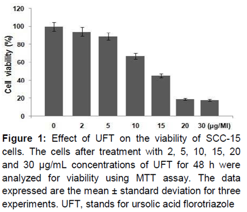
Results: Treatment of SCC-15 cells with UFT for 48 h significantly reduced cell viability in a dose-dependent manner. At 20 µg/mL concentration of UFT, SCC-15, cell viability was reduced to 19 % compared to 100 % in the untreated cells (p = 0.0002). UFT treatment enhanced the proportion of apoptotic cells which was evident from YO-PRO-1 staining. In UFT-treated cultures, the population of cells in sub-G1 phase increased to 38.54 % compared to 7.32 % for control after 48 h. ex
Conclusion: UFT treatment inhibits viability and induces apoptosis in squamous cell carcinoma cells through suppression of Fas ex
Introduction
Squamous cell carcinoma is the commonly observed malignant tumor of mucosa lining of the oral cavity. In Korea squamous cell carcinoma accounts for more than 90 % of the detected malignant tumors of the oral cavity [1]. Despite advancement in the fields of chemotherapy, radiation therapy and surgery, the prognosis of oral squamous cell cancer patients remains poor [2]. The five year survival rate of oral squamous cell cancer patients has been observed to be 50 % [2]. Taking into consideration the poor survival rate, the discovery of drugs for the treatment of patients with squamous cell carcinoma is desperately required.
Apoptosis, the programmed and controlled process of cell death is regulated by both internal as well as external signals. It plays an important role in the development and maintenance of multicellular organisms by eliminating unwanted cells from the body [3]. Apoptotic cells are characterized by morphological alterations including, decreased cell and nucleus sizes, blebbing of plasma membrane, condensation of chromatin and DNA fragmentation [4]. Induction of apoptosis in carcinoma cells by treatment with chemotherapeutic agents is a promising strategy for cancer treatment [5,6].
Ursolic acid (UA), belonging to the terpenoid family of natural products is a pentacyclic molecule abundantly present in the plant kingdom [7-10]. It is a biologically active compound and has medicinal importance in traditional Chinese medicine for the clinical treatment of various diseases. UA exhibits a broad spectrum of activities including, as anti-inflammatory, antiviral, antioxidant, hepatoprotective, cytotoxic, antitumor, anti-angiogenesis, and anti-metastatic activities [11]. It has also been demonstrated that UA causes inhibition of colon carcinoma cell proliferation and induces apoptosis [12,13-15].
It was reported that the triazole analogs of UA are more potent compared to the parent compound. In the present study, the effect of ursolic acid floro triazole (UFT) on oral squamous cancer was investigated. The results revealed a significant reduction in cell viability, induction of apoptosis, cell cycle arrest and reduction in tumor weight and volume in vivo.
Methods
Drug and chemical
Ursolic acid florotriazole (UFT) was synthesized using a previously reported procedure from the parent ursolic acid using reported procedure [13]. UFT was dissolved in DMSO to prepare the stock solution and stored at -20 oC.
Animals
Female C3H/HeN mice (8 weeks old) were purchased from the Laboratory Animal Center, Third Military Medical University (Chongqing, China). The animals were housed under 12 h light and dark cycle in pathogen‑free environment, with access to water and food ad libitum.
All the animal experiments were performed according to the Kyung Hee University Institutional Animal Care and Use Committee guidelines. The study was approved by the ethics committee of Kyung Hee University Institutional Animal Care and Use (approval reference no. 012-47-KHI.
Cell culture
SCC-15 human squamous cell carcinoma cell line was purchased from The Cell Bank of Type Culture Collection of Chinese Academy of Sciences, Shanghai Institute of Cell Biology (Shanghai, China). The cells were maintained at 37 ˚C in an atmosphere of 5 % CO2 in RPMI-1640 medium (Thermo Fisher Scientific Inc., Waltham, MA, USA). The study was approved by the Ethics Committee of Kyung Hee University (Yongin, Korea).
MTT assay
Into the 96-well flat bottom multiplates (BD Falcon, Franklin, NJ) cells at a density of 2.5 x 10⁵ cells per well were distributed. To each of the well different concentration of UFT was added and incubated for 48 h. MTT (Sigma-Aldrich, St. Louis, MO, USA) solution (10 μL) was added to each well and the plates were incubated for 4 h at 37 oC in 5 % CO2 incubator. Formazan crystals were dissolved by adding 0.04 N HCl in 2-propanol (100 μL). The microplate reader (MPR-A4i; Tosoh Corporation, Tokyo, Japan) was used to measure absorption at 570 nm. All the measurements were carried out in triplicate.
Confocal microscopy
SCC-15 cells at a density of 2 x 10⁶ cells per well were distributed onto 6-well plates (Nunc A/S) containing RPMI-1640 supplemented with 2 % FBS. Following 24 h incubation, the cells were treated with a range of UFT concentrations (2, 5, 10, 20 and 30 µg/mL). After 48 h, the cells were washed twice with ice-cold PBS, treated with 20 µL PBS and subsequently stained for 30 min with 0.2 µM YO-PRO-1 (Molecular Probes Inc., Eugene, OR, USA) dye. The confocal laser microscope (LSM 510 Meta; Carl Zeiss, Oberkochen, Germany) with a 490 nm argon laser was used to record the fluorescence of the cells and capture the images.
Cell cycle analysis
In 100 mm culture dishes, SCC-15 cells were distributed at a density of 2 x 10⁵ cells per well and incubated with various concentrations of UFT for 48 h. The cells were then harvested, centrifuged for 10 min at 500 x g and subsequently rinsed in ice‑cold PBS. The cells were fixed in 70 % ethyl alcohol and stored for 2 h at -20 ˚C. Followed PBS washing, the cells were put in 200 µL PI/RNase Staining Buffer (BD Biosciences) for 1 h under dark conditions. FACSVantage SE flow cytometry system and CellQuest program (BD Biosciences) were used for the analysis of DNA content of the stained cells.
Reverse transcription (RT)‑PCR
The cells were cultured in medium containing UFT followed by RNA extraction using Isogen systems reagents (Nippon Gene Co., Ltd., Tokyo, Japan). Following denaturation by heating at 60 ˚C for 30 min using 500 pmol oligo(dT) primer, RNA was reverse-transcribed to cDNA. The reverse-transcription solution comprised of Moloney murine leukemia virus reverse transcriptase (Invitrogen Japan K. K., Tokyo, Japan), 75 mm KCl, 50 mm Tris-HCl (pH 8.3), 3 mm MgCl2, 0.01 M DTT, 0.5 mm of each dNTP and 16 units RNasin® (Promega Corporation, Madison, WI, USA). Quantitative PCR and BI Prism 7700 Sequence Detector (TaqMan; PE Biosystems Japan, Tokyo, Japan) were used for the amplification of cDNA. The reaction was performed in a 50 µL reaction mixture containing 200 nM forward and reverse primers, 100 nM probe specific for the targeted cDNA and TaqMan Universal Master mix (PE Biosys- tems Japan). The data were examined using Sequence Detection software (PE Biosystems Japan). The primers and probes for amplifying Fas were purchased from PE Biosystems Japan. Target gene expression levels were standardized to the internal standard gene expression levels. The relative expression levels of Fas in the UFT-treated cells were compared with the levels in the untreated cells. Enhanced chemiluminescence system was used for visualization of immunoreactive bands.
Tumor growth in in vivo SCC-15 animal model
To establish an in vivo SCC-15 animal model, 2 x 10⁵ SCC-15 cells in 100 µL PBS were subcutaneously injected into the right flank of the C3H/HeJ mice. After five days, when the tumors formed visible masses, the animals were divided into two groups, each containing 5 mice. The animals in treatment group were injected intraperitoneally 20 mg/kg body weight of UFT daily in the morning for 15 days whereas the animals in control group received PBS. On day 30 following carcinoma cell administration, the animals were euthanized using CO2 and the tumors were extracted and weighed. The volume of the tumor was determined after measurement of tumor dimensions using a caliper.
Statistical analysis
For the purpose of statistical analysis, Mann-Whitney U test was used. Student’s t-test was used for statistical analysis of the effect of UFT on cancer cell proliferation and the alteration of mRNA expression in vitro. P < 0.05 was considered to indicate a statistically significant difference.
Results
Suppression of SCC-15 cell viability by UFT
The effect of various concentrations (0, 2, 5, 10, 15, 20 and 30 µg/mL) of UFT on the viability of SCC-15 cells was evaluated by MTT assay. The results revealed a concentration dependent reduction in the viability of SCC-15 cells on treatment with UFT for 48 h. Among the various concentrations of UFT tested, the reduction in cell viability was maximum at 20 µg/mL concentration after 48 h. At 20 µg/mL concentration of UFT, SCC-15 cell viability was reduced to 19 % compared to the untreated cells (p = 0.0002, ).
Induction of apoptosis in SCC-15 cells by UFT
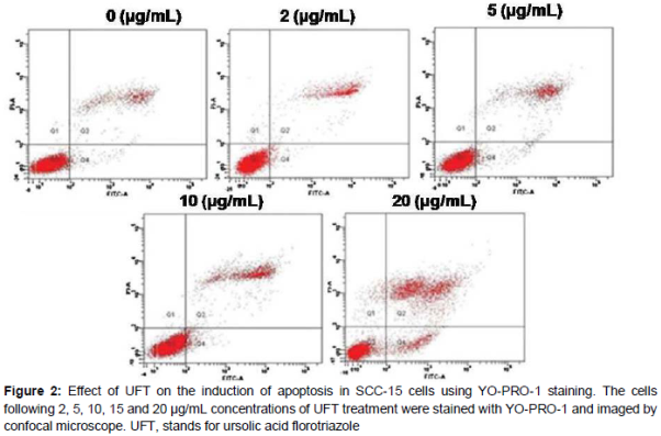
Induction of apoptosis in SCC-15 cells by UFT treatment was examined using YO‑PRO‑1 dye followed by confocal microscopy. The results showed a significant increase in the proportion of apoptotic cells with increase in the concentration of UFT from 5 to 20 µg/mL ().
UFT increased the number of cells in sub-G1 phase
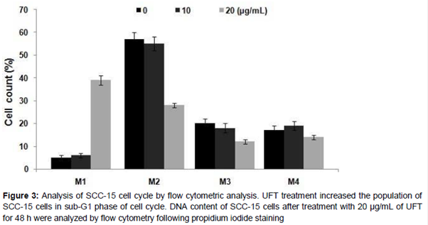
Analysis of cell cycle using flow cytometry after PI staining revealed a significant increase in the proportion of cells in sub-G1 phase (A). UFT treatment at 20 µg/ml increased the proportion of cells in sub-G1 phase to 38.54 % compared to 7.32 % in the control cells after 48 h.
Effect of UFT treatment on the expression of Fas gene in SCC-15 cells
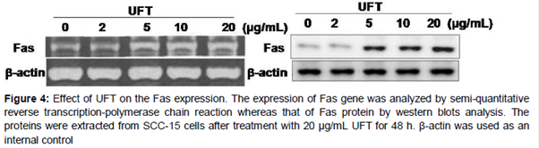
Expression of Fas gene in SCC-15 cells was determined by RT-PCR. The results showed a significant increase in the expression of Fas on treatment with 20 µg/mL concentration of UFT compared to that of untreated cells (). The increase in the expression of Fas by UFT treatment was further confirmed using western blot analysis (). These data indicate that UFT increases the expression of Fas in SCC-15 cells.
UFT treatment inhibits tumor growth in an SCC-15 animal model
The antitumor activity of UFT treatment was examined in an SCC-15 animal model using C3H/HeJ mice. At five days after subcutaneous implantation of SCC-15 cells, the mean tumor volume was ~10 mm3. In the control (PBS-treated) group, the tumors grew rapidly and reached an average volume of 501 ± 43.9 mm3 (mean ± SD) on day 14 after inoculation with SCC-15 cells (A). The mean size of the primary tumor in animals treated with 10 mg/kg UFT was determined to be 202 ± 20.2 mm3 at 14 days. Similarly, the mean tumor weight for the animals treated with 10 mg/kg UFT 46.2 % of that in the control group (B). Together, these results indicate that UFT inhibited tumor growth in the SCC-15 animal model.
Discussion
The present study demonstrates effect of UFT on the cell viability, induction of apoptosis and the mechanism underlying the in vitro inhibition of oral squamous cancer cells as well as in vivo mouse model. It was observed that UFT treatment inhibited SCC-15 cell viability, induced apoptosis, and increased the proportion of cells in sub-G1 phase. In addition, the tumor volume and weight in the UFT-treated mice were also reduced significantly compared to the untreated mice.
Inhibition of the viability of carcinoma cell lines by chemotherapeutic agents can be a promising strategy for the treatment of cancers. The results from the present study showed that UFT exhibited a concentration dependent inhibitory effect on the viability of SCC-15 cells following exposition for 48 h. UFT treatment increased the proportion of apoptotic cells significantly compared to the untreated cells. Analysis of cell cycle revealed a marked increase in the population of cells in sub-G1 phase with increase in the concentration of UFT to 20 µg/mL. It is reported that Fas exhibits a vital role in the induction of cell apoptosis in various types of carcinoma cells through the caspase activation pathway [16]. UFT treatment for 48 h caused enhancement in the expression of Fas gene in SCC-15 cells significantly compared to the untreated control cells. Thus, UFT treatment leads to apoptosis in SCC-15 cells via Fas signaling pathway activation. In the present study, the antitumor activity of UFT was also investigated in a SCC-15 animal model using C3H/HeJ mice. Reduction in the tumor volume and weight has been observed in various types of mouse xenograft models including, renal and prostate carcinoma [17,18]. In the present study, it was observed that UFT treatment caused a significant suppression in the volume and weight of tumor in SCC-15 animal model.
Conclusion
The results from the present study reveal that UFT inhibited cell viability, induced apoptosis of oral squamous cell cancer cells, and suppressed tumor growth in vivo through Fas signaling pathway. Thus, UFT is a potent candidate for the treatment of squamous cell cancer.
References
Archives


News Updates